
Table of Contents
Understanding the differences between Simple and differential staining methods is crucial for microbiologists, as each serves a unique purpose in research, diagnostics, and education.
Staining is a cornerstone in microbiology, enabling scientists to visualize and study microorganisms under a microscope. Due to their transparent and colorless nature, many microorganisms would remain invisible without staining. Staining enhances visibility and provides valuable information about microbial cells’ structure, shape, and characteristics.
This article explores the principles, processes, and applications of simple and differential staining, highlighting their significance in studying microorganisms.
What is Simple Staining?
Simple staining is a basic microbiological technique that uses a single dye to color microorganisms, making them more visible under a microscope. This method highlights the overall structure and morphology of microbial cells without providing detailed information about specific cellular components or differences between types of cells.
Purpose
The primary purpose of simple staining is to observe the basic morphology of microorganisms, including their:
- Shape: Whether they are spherical (cocci), rod-shaped (bacilli), spiral (spirilla), or other forms.
- Size: Determining relative dimensions of cells.
- Arrangement: Observing how cells group, such as in chains, clusters, or pairs.
This technique is particularly useful for preliminary studies where a quick and general observation is needed.
Simple Staining Procedure
- Smear a small microorganism sample onto a glass slide and air-dry.
- Heat fix the slide to adhere the cells to the surface and kill the microorganisms.
- Apply a single stain, such as methylene blue, eosin, crystal violet, or safranin, to the smear for a specific period.
- Rinse the slide gently with water to remove excess stain.
- Examine the stained sample under a light microscope,the cells appear prominently against the background.
Examples
Some common dyes used in simple staining include:
- Methylene Blue: Highlights bacterial cells and is commonly used in bacteriology.
- Safranin: Produces a reddish-pink stain and is often used as a counterstain but works well for simple staining.
- Crystal Violet: A vibrant purple stain that provides excellent contrast.
Advantages
Simple staining offers several benefits:
- Quick and Easy: The process is fast and requires minimal reagents and steps.
- Cost-Effective: Single dyes are inexpensive and widely available.
- Ideal for Beginners: The technique is straightforward, making it suitable for students and novice microbiologists.
- Basic Visualization: Provides sufficient information to identify cell morphology and arrangement for initial studies.
Simple staining provides a clear, enhanced view of microbial cells, serving as a foundational tool for further microbiological analysis and more complex staining techniques.
What is Differential Staining?
Differential staining is an advanced microbiological technique that uses two or more dyes to differentiate between types of microorganisms or distinct cellular structures. This method allows microbiologists to identify specific characteristics of cells, such as differences in cell wall composition or the presence of unique cellular components.
Purpose
The main purpose of differential staining is to provide detailed information about microorganisms, aiding in their classification and diagnosis. This technique is particularly useful for:
- Identifying Types of Bacteria: Distinguishing between Gram-positive and Gram-negative bacteria.
- Studying Cell Wall Structures: Highlight variations in cell wall composition, such as those in acid-fast bacteria.
- Detecting Special Features: Observing specific cellular components, such as spores, capsules, or flagella.
Differential staining is critical in clinical diagnostics and research by offering precise insights into microbial characteristics.
Differential Staining Procedure
- Apply a primary dye to the sample to color all cells or components. For example, crystal violet is the primary stain in Gram staining.
- Use a decolorizing agent (e.g., alcohol or acid-alcohol) to remove the primary stain from certain cells or structures selectively. This step highlights differences based on the cell’s physical or chemical properties.
- Apply a secondary dye (counterstain) to color the decolorized cells, providing contrast. For instance, safranin is used in Gram staining to stain Gram-negative bacteria pink.
Examples
Differential staining techniques include:

- Differentiates bacteria into Gram-positive (purple) and Gram-negative (pink) based on their cell wall composition.
- Acid-Fast Staining:
- Identifies acid-fast bacteria (e.g., Mycobacterium tuberculosis) that retain the primary stain (carbol fuchsin) despite decolorization with acid-alcohol.
- Endospore Staining:
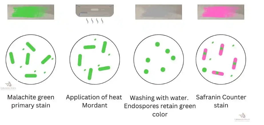
- Highlights bacterial spores, which appear green after staining with malachite green and counterstaining with safranin.
Advantages
Differential staining provides several key benefits:
- Diagnostic Value: Essential in identifying pathogenic microorganisms in clinical samples.
- Detailed Information: Reveals structural differences and special features not observable with simple staining.
- Broad Applicability: Used in bacteriology, mycology, and other fields of microbiology.
- Research Utility: Facilitates the study of microbial diversity and cellular adaptations.
Differential staining is an indispensable tool in research and diagnostic microbiology, offering a deeper understanding of microbial structures and properties.
Key Differences Between Simple and Differential Staining
Simple staining and differential staining are fundamental techniques in microbiology, but they serve different purposes and involve distinct processes. The table below highlights their key differences:
| Aspect | Simple Staining | Differential Staining |
|---|---|---|
| Definition | Uses a single dye to color all microorganisms uniformly. | Uses multiple dyes to distinguish between different types of microorganisms or cellular structures. |
| Number of Dyes Used | One (e.g., methylene blue, safranin, crystal violet). | Two or more (e.g., crystal violet and safranin in Gram staining). |
| Complexity of Process | Simple with minimal steps. | More complex, involving multiple steps like decolorization and counterstaining. |
| Purpose and Information Obtained | Provides basic information about cell shape, size, and arrangement. | Provides detailed information, such as cell wall structure, presence of spores, or acid-fastness. |
| Examples of Use | Preliminary observations in bacteriology or basic morphology studies. | Diagnostic applications (e.g., identifying Gram-positive or Gram-negative bacteria) and detailed structural analysis. |
Simple staining is ideal for quick, general observations, while differential staining is crucial for more detailed and diagnostic purposes.
Applications in Microbiology
Staining techniques play a vital role in microbiology, aiding researchers and clinicians in visualizing microorganisms and understanding their characteristics. Depending on the level of detail required, simple and differential staining have distinct applications.
When to Use Simple Staining
- General Observations: It provides a quick overview of microbial shape, size, and arrangement, making it ideal for preliminary studies or when detailed analysis is unnecessary.
- Teaching and Training: Often used in educational settings to introduce students to basic microscopy and staining techniques.
- Routine Laboratory Work: For rapid visualization of microorganisms in a sample where identification is not the primary objective.
When to Use Differential Staining
- Diagnosing Diseases: Techniques like Gram staining classify bacteria in clinical samples, helping identify pathogens responsible for infections. For example:
- Identifying Gram-negative bacteria like Escherichia coli in urinary tract infections.
- Detecting acid-fast bacteria like Mycobacterium tuberculosis in suspected tuberculosis cases.
- Classifying Microorganisms: Provides critical insights into bacterial taxonomy, distinguishing between Gram-positive and Gram-negative bacteria or acid-fast and non-acid-fast organisms.
- Research on Cellular Structures: Techniques like endospore staining study bacterial adaptations and survival mechanisms.
Examples of Research and Clinical Applications
- Investigating microbial diversity in environmental samples using differential staining.
- Studying bacterial spore formation and survival strategies with endospore staining.
- Examining biofilms and cell wall properties in Gram staining experiments.
- Diagnosing bacterial infections through Gram staining in medical laboratories.
- Detecting acid-fast pathogens in respiratory samples during tuberculosis testing.
- Identifying fungal spores and yeasts using specific staining techniques.
Conclusion
Staining techniques are fundamental to microbiology, offering insights into the often invisible world of microorganisms. Simple and differential staining are two key methods that serve distinct purposes. While simple staining provides a quick and efficient way to observe the basic morphology of microorganisms, differential staining offers a deeper understanding of their structural and functional characteristics, making it indispensable for classification and diagnostics.
This article highlights the differences between these techniques and emphasizes the importance of choosing the appropriate method based on the level of detail required. Whether used for basic observations, teaching, advanced research, or clinical diagnostics, both staining techniques play complementary roles in the study and application of microbiology. Understanding and mastering these methods is essential for microbiologists to unlock the secrets of microbial life.